
A newly developed technique excels at capturing 3-D images of cells in their natural environments.Researchers have made an imaging breakthrough that lets them capture 3D footage of cells doing their thing inside the body — and it may look nothing like what you imagined.
source/image(PrtSc): HHMI Howard Hughes MI
The cells on the surface of the fish act like water on a car windshield, obscuring and scattering any light that tries to penetrate them. The further you look into the organism, the worse the distortion becomes.
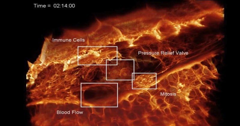
Immune cells within the perilymphatic space of the inner ear of several zebrafish embryos 80 hpf showing: MIP view of two immune cells (orange), one of which has ingested dextran particles (blue).
Advertisement
Before and after AO plus deconvolution for 438 time points at 13 sec intervals; volume rendered view in another embryo. Showing a migrating immune cell and a dividing endothelial cell; and tracking of the position and velocity of an immune cell in a third embryo (c.f., Fig. 6E,F, figs. S13-15).


